TELOCYTES: A NOVEL PLAYER IN PRESERVING THE HOMEOSTASIS OF RAINBOW TROUT Oncorhynchus mykiss INTESTINAL MUCOSA
Intestinal epithelium undergoes a rapid, constant, and complete renewal, driven by a specialized population of intestinal stem cells (ISCs) that are housed in well-defined niches made of epithelial and stromal cells. We previously described the spatial organization of the rainbow trout (RT) ISCs, identifying the epithelial and stromal niche components in this species. Here, we expand that knowledge focusing on a newly identified stromal population known as telocytes that has recently been identified as an active player of the intestinal mucosa.
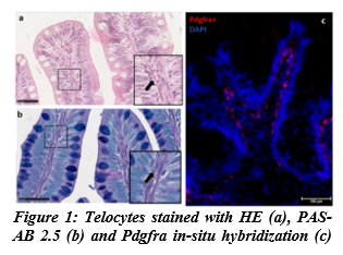
Segments of proximal and distal intestine were collected from 5 female rainbow trouts (Oncorhynchus mykiss) weighing around 250g. Sections were stained with hematoxylin-eosin (HE), Periodic Acid Schiff-Alcian Blue at pH 2.5 (PAS-AB 2.5), Mallory's triple stain, Crossman's trichrome, toluidine blue and silver nitrate. Samples were also subjected to in situ hybridization for pdgfrα, foxl1 and lgr5, that are known to selectively identify intestinal telocytes and telocytes' subsets. Finally, small fragments of proximal and distal intestine were examined with transmission electron microscopy (TEM), looking for telocytes peculiar ultrastructural characteristics.
Slender, elongated cells distributed in the peri-epithelium region around the folds base and along their whole length, were detected in both gut regions. These cells showed affinity for all the histochemical staining and expressed pdgfrα (fig. 1). TEM further confirmed their identity as telocytes. They created a network extending from the folds base to their apex. Some of them expressed also foxl1, while others expressed lgr5, indicating the presence of different subsets. Telocytes positive to both pdgfrα and foxl1 were located around the fold base close to sox9+ cells, that we previously identified as the real rainbow trout ICS. This suggests that some telocytes may support the epithelium renewal and homeostasis. Telocytes positive to foxl1, lgr5 and pdgfrα were selectively located in the folds apex, implying a different role aimed at the maintenance of the tip epithelium.
Our data showed that two different telocyte subpopulations are present in the RT intestinal mucosa: one forms a peri-cryptal signaling source, possibly promoting and modulating ISC proliferation. The other, located at the fold apex, may be involved in modulating cell differentiation and anoikis. Taken together our results suggest that telocytes could represent relevant targets for the morphological evaluation of diet effects on RT gut health
Supported by the European Union's Horizon 2020 research and innovation programme under grant agreement No 828835 and by the AGER-2 Sushin project, cod.2016-0112.